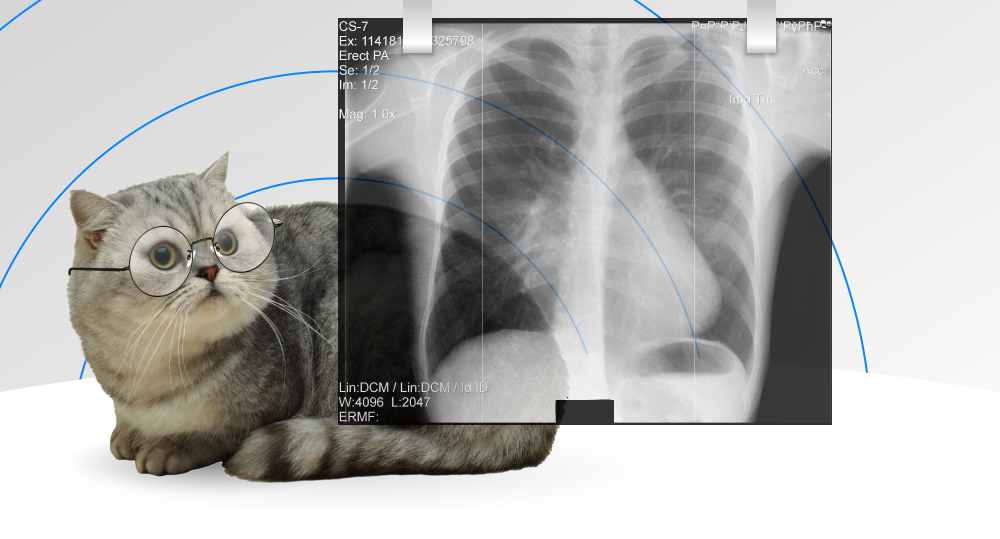
The raging storm of COVID-19 waves keeps on testing our healthcare systems. There has never been a greater demand for modern technological solutions used for professional diagnostics, treatment, and prediction of health conditions and specialized support and assistance of continuously overworked medical staff. According to The Lancet journal, machine learning in radiology is one of the most influential fields for innovations in the sphere. They believe that machine learning algorithms, neural networks, and deep learning have a high potential for the new era of advanced medical help.
We have chosen for you trends and prospects in implementing the latest achievements in machine learning that are transforming healthcare as we speak.
WRITTEN BY:
Aliaksei Sliborski
Senior Software Developer, Qulix Systems
Contents
Machine Learning: Radiology Research Transformed
With the pandemic still affecting the planet's population, the strong demand for qualified diagnostic specialists, especially radiologists, is at the highest rate. Since the primary methods for detecting severe coronavirus cases are chest X-rays and CT images, specialists have to process hundreds of such photos every day. Big data on COVID-19 has become available for machine learning and neural networks, applying deep learning methods and algorithms. That promises critical support in diagnosis. We are sure that implementing machine learning in radiology is crucial for overcoming pandemic outbreaks. Moreover, it will transform various health industry processes to achieve higher service levels.
If we look at the radiology department, it has always been on the frontline of technology in healthcare. In almost all medical fields, innovative approaches to medical imaging methods evolved into new interpretation, classification, and implementation technologies. No wonder that the opportunities for artificial intelligence, and machine learning particularly, became so promising.
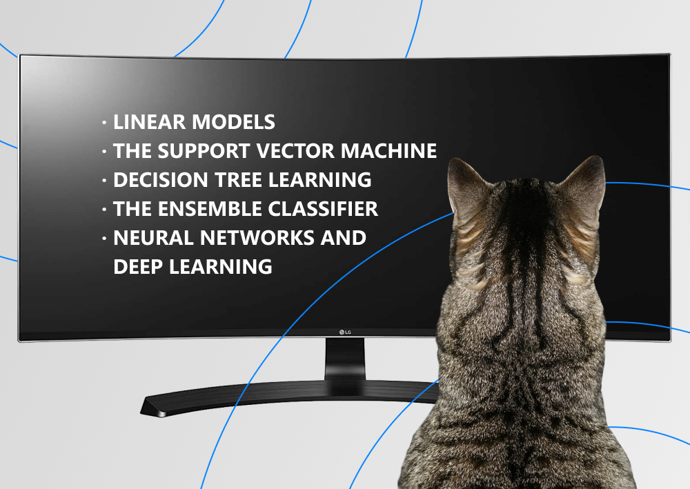
Several machine learning methods have proved their effectiveness for application in radiology:
- linear models are the most common ones for radiology are logistic regression, linear regression, and Fisher's linear discriminant (LDA);
- the support vector machine is a set of kernel-based supervised learning techniques used for regression and classification; it finds an optimum hyperplane for linear separable patterns;
- decision tree learning is one of the top classification approaches in machine learning used for forming random forests for classification and prediction in the analysis of radiological images;
- the ensemble classifier is a combination of multiple classifiers that leads to final classification by bagging and boosting;
- neural networks and deep learning can pick specified features out of the data to detect and classify them.
Machine learning algorithms, detection, classification, and prediction solutions can empower physicians to provide patients with advanced, more innovative care. We will enlighten the main routes for developing ground-breaking computer-assisted healthcare.
Machine Learning Image Interpretation in Radiology
The first and the most obvious use case of machine learning is interpreting images produced for examinations, diagnostics, surveillance, and health condition research. According to the recent study by Frost & Sullivan, the global medical imaging informatics market is rapidly growing. It is estimated to reach $10.4 billion by 2025 with a CAGR of 3.5% since 2019. Medical imaging informatics is the top moving gear in treating and managing illnesses.
The most significant revenue contributors to innovations are forecasted to be artificial intelligence and machine learning in radiology. They have a considerable potential to identify and classify compound patterns from a range of radiological images — X-rays, CT scans, MRIs, and PET scans. Machine learning algorithms in radiology can become one of the main parts of future decision-making in different healthcare fields.
Among the leaders in developing and implementing machine learning techniques, we have chosen some peculiar and innovative brands for you.
Arterys is a company that originated in Stanford University's labs. In 2017 they began training their machine learning model to detect heart abnormalities in radiologic images. Now their models can deal effectively with different organs and systems of the human body. The distinctive feature of ArterysAI is in using cloud computer processing to make all the data available despite the location. The sensitive PHI data is cut out of the images before uploading to the cloud. After the analysis, the PHI, stored on hospitals’ internal servers, is applied to the results; thus, ArterysAI never gets access to it.
MaxQ AI sees its mission in reinventing patient diagnosis through artificial intelligence, machine learning, and deep learning. The MaxQ AI software ACCIPIO is based on machine learning and computer vision technology and focuses on stroke and head trauma care. It delivers precise, seamless, and secure assessments and aims to prevent more than 40 thousand deaths from misdiagnosis a year.
Zebra is an Israeli company that has brought machine learning in medical imaging interpretation to the next level. They started their work back in 2014 with two years of careful research of a complex of data: different modalities of radiology images, lab analyses, doctors' diagnoses, and clinical outcomes. They built Med's Imaging Analytics Engine, which is integrated into hospitals' existing software workflow environment and provides a secure and robust computer-assisted solution for early detection and treatment of diseases to provide optimal care pathways for patients.
Convolutional Neural Networks for Image Classification
The promising method for radiology is deep learning, and particularly convolutional neural networks (CNN) applied for image classification or, in plain English, detecting normal and abnormal conditions of human organs. For example, the CNN method proved its effectiveness for identifying pneumonia cases with 91% accuracy. The approach uses 5.5 thousand images of lung X-rays — about 3 thousand with lung opacity and 1.5 thousand normal ones. The next important step is Image Augmentation to increase dataset variation for deep learning.
After that, the convolutional neural network is applied. The CNN architecture uses layers of convolution to receive the input and then transforms the data of the image as input for passing it to the next layer. Each layer uses several filters for detecting edges, curves, objects, textures, and other patterns. The filters, respectively, are image kernels, a matrix of 3x3 or 4x4 applied to an image on the whole.
The callback list has to be defined to fit the model. It will help avoid overfitting and assigning class weights to learn from all classes. And the final stage is to run the CNN model and then detect the images and interpret the results.
Machine Learning: Radiology beyond Image
Are there any other prospects for artificial intelligence and machine learning in radiology? We can suggest several aspects where implementing computer science can effectively empower the workflow for radiologists.
Image Display and Study Protocols Upgrade
Radiological medical imaging produces metadata, and it is crucial to use the optimal display method for studies to interpret them appropriately. For example, there is a complex set of dozens of medical images in different anatomic planes during an MRI. In addition, there can be images from various tools available. Using machine learning algorithms to display all the studies accurately increases radiologists' productivity.
One more possibility for machine learning to effectively reduce the amount of routine workflow is to assist in the appropriate creation of study protocols. AI is applied to review and arrange all the patient information stored in an electronic health record: relevant prior images, test results, and radiology reports. Such software becomes a handy time-saving tool in clinical practice.
Image Quality Improvement
Furthermore, machine learning can also immensely transform the technique of taking medical images. It has always been a big concern for radiologists to reduce the radiation dose during medical imaging, resulting in poor image quality. Implementing deep learning techniques eliminates image noise and helps get high-quality images while using ultra-low-dose CT protocols. However, MRI imaging has the prospect of reducing the radiation dose by rapid acquisition protocols. Deep learning reconstructs anatomic MRIs with limited raw data and thicker MR scans and halves the acquisition time. These solutions provide optimization of tools used in this very costly healthcare field.
Scanner Usage Optimization
There are also several other ways to reduce the expenses. One of them is to schedule staff for shifts. It depends on a vast set of requirements — the day of the week and time of the day, exam complexity and volume of studies, the department of the hospital, and clinical practice patterns. Machine learning can optimize staffing and increase profitability and cost reduction. It can also provide solutions for patient "no-shows" by implementing algorithms based on numerous factors such as the time of visits and patient demographics. All that highly improves cost-effectiveness.
Prediction Models
Radiological medical images, descriptions, and interpretations are essential for detecting disease and predicting future health conditions. Machine learning algorithms applied to health data sets can create precise prediction models, e.g., tolerance to medication, tumor response to chemotherapy, or survival odds of operative treatment. That is one more important support line for medical professionals.
Quite a promising perspective for transforming radiology and healthcare in general? Not that simple.
Obstacles to Overcome
We see that machine learning has considerable potential in radiology. Nevertheless, it is still not a substitute for the clinician and only an assistant. There are a lot of limitations to implementing these technological solutions in clinical practice in the current stage of development. The main obstacles that researchers should try to overcome are pretty serious.
Learning Methods
The first limitation is creating applications of machine learning for radiology. Most of the studies are based on previously available decisions that radiologists made and supervised learning methods developed. Will we ever be able to rely on diagnosis by machine learning techniques in actual medical practice without any additional supervision? That is an earnest question by now.
Data Limits
One more roadblock for machine learning in radiology lies in sensitive patient data. Robust machine learning models require large amounts of datasets for analysis. Limited access to patients' information due to regulations and different clinical practices still leads to a limited number of relevant cases available for research.
Accuracy
The level of accuracy is a very significant obstacle. The price of even the tiniest incorrectness in this sphere can be too high. The algorithms are expected to provide a 100% accuracy rate to have no doubts in implementing into actual healthcare practice. Nevertheless, that is still a future goal for researchers.
Takeaways
In a nutshell, machine learning has enormous potential for transforming patient care and improving it dramatically. Essentially, machine learning algorithms can solve so many problems alongside medical image interpretation for radiology. We are sure that the careful symbiosis of radiology experts and improved practical applications of machine learning will undeniably lead to the next level of healthcare for the whole world.
Our Artificial Intelligence cluster has the tremendous experience to extend your capabilities through the latest technological innovations. You are welcome to contact our professionals to get the best support for your ideas.

Contacts
Feel free to get in touch with us! Use this contact form for an ASAP response.
Call us at +44 151 528 8015
E-mail us at request@qulix.com

